HIV-1
Reverse Transcriptase
David Marcey
© 2006
I.
Subunit structure
II. Nucleic acid interactions
III. RT Inhibitors
IV. References
Directions
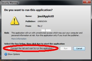
If
prompted, allow your browser to view blocked content. The OMM
now uses the Signed Jmol Applet. No files from this site can
damage your computer. Just check the appropriate box and then
click run.
This
exhibit displays molecules in the left part of the screen, and
text that addresses structure-function relationships of the
molecules in the right part (below). Use the scrollbar to the
right to scroll through the text of this exhibit.
To evoke renderings of the molecule that illustrate particular
points, click the radio buttons:
To
load/reset the molecule, use these buttons:
If
you resize your browser window, simply refresh the page in order
to restore proper viewing.
|
I.
Subunit structure
The enzyme reverse transcriptase
(RT) is used by retroviruses to transcribe their single-stranded RNA
genome into single-stranded DNA and to subsequently construct a complementary
strand of DNA, providing a DNA double helix capable of integration
into host cell chromosomes. Functional HIV1-RT is a heterodimer containing
subunits of 66 kDa (p66) and 51 kDa (p51).
p66 contains two the two domains responsible
for the two catalytic activities of RT, the N-terminal polymerase
domain and the C-terminal RNase H domain.
The polymerase domain catalyzes polymerization
of DNA in a primer strand complementary to a template strand of RNA
(see below). The RNase H domain catalyzes
the degradation of the RNA template (see below).
p51 is processed by proteolytic cleavage
of p66 and corresponds to the polymerase domain
of the p66 subunit.
The polymerase domain of p66 includes
three subdomains that can be described as the fingers,
palm, and thumb
of a clasping right hand:

The
hand subdomains
serve to clasp the RNA-DNA duplex in the process of RNA directed DNA
polymerization. The fingers and thumb
domains are the walls of a nucleic acid binding cleft, with the palm
subdomain seving as a base containing the DNA polymerase active site
(see below).
The connection subdomain
connects the hand subdomains
of the polymerase domain and the RNase H domain
of p66, which provides the ribonuclease activity of HIV-RT, digesting
the RNA template after a DNA copy is polymerized.
The p66 palm
and
connection
subdomains contain
three-stranded beta sheets with alpha
helices on one side. The thumb
subdomain comprises
three alpha
helices.
Interestingly, although p66
and p51 are identical in their primary
amino acid sequence (except for length) and they share similar subdomain
structures, they are topologically quite distinct.
For example, three
catalytic, aspartate residues
from the palm subdomain are exposed in
the nucleic acid binding cleft of p66,
but are buried in p51, which lacks a
discernable cleft.
Another striking difference between
the two subunits is the orientation of the connection
subdomain; in p51 it is tucked into a
central position and contacts all of the other subdomains, but in
p66 it contacts only RNase
H and the thumb.
A question
arises as to why HIV has evolved a heterodimer in which the smaller
subunit (p51) is a cleavage product of the larger. One speculation
(e.g. Kohlstaedt, et al., 1992) is that the selection for streamlined
genomes in retroviruses has forced the evolution of different protein
subunits encoded by the same gene. In the case of HIV-RT, subunits
with different structural and functional properties can be produced
by proteolytic cleavage of one of two initially identical subunits.
return
to beginning of the exhibit
II.
Nucleic Acid-RT Interactions
HIV-1 RT contains an ~60 Angstrom groove between the
polymerase and RNase
active sites. The connection
subdomains of both p66 and p51
form the floor of this groove.
The template-primer
nucleic acid duplex fits into this groove and is cradled by the hand
subdomains
of p66, which form a nucleic acid binding
cleft that includes the polymerase active site (see below).
Numerous
residues from the fingers, palm,
thumb, and connection
subdomains and the RNase
H domain contact the nucleic acid backbone. Residues from one
helix of the thumb subdomain contact
bases directly.
The
few, direct contacts between the nitrogenous bases of the template-primer
double helix and
RT residues are mostly van der Waals interactions. These include minor
groove base interactions with thumb residues
of helix H, palm
residues near the primer strand 3' terminus, and an RNase
H domain residue. Hydrogen bonding between tyr183
and a G in the minor groove is observed.
Focusing now on the polymerase active site, the incoming nucleotide
(in this case, dTTP) is positioned to
be added to the growing primer strand.
Nucleophillic attack on the alpha phosphate
of the incoming nucleotide by the 3' oxygen
on the 3' carbon of the primer terminus will produce the covalent
linkage of the new nucleotide to the primer strand The two remaining
phosphates of dTTP
will form a leaving group. The dTTP is
positioned by hydrogen bonding with a complementary base in the template
strand, and by
interactions with Mg++ ions
and residues of the palm and fingers
subdomains. Three catalytic aspartate
residues from
the palm subdomain are involved in coordinating
the Mg++ ions. In a well studied,
two-metal mechanism found in numerous other polymerases, these ions
serve two key functions: 1) stabilizing the ionized form of the 3'
oxygen (O-), increasing its nucleophilicity and leading to
the attack on the alpha phosphate; 2)
stabilizing negative charges on the diphosphate
leaving group.
*
At left are the
superimposed protein backbones of unliganded
HIV-1 RT (Rodgers, et al., 1995) and the liganded
form of the enzyme (Huang, et al., 1998), bound to a double helical,
nucleic acid, template-primer substrate (not shown). Although the
backbones are largely congruent in the RNAase H domains and the connection
subdomains of p51 and p66, the arrangement of the hand subdomains
of p66 changes upon binding nucleic acid.
These changes are readily observed by viewing the two forms of the
enzyme sequentially. Template-primer binding causes the fingers subdomain
to curl toward the palm in the liganded
enzyme, relative to the unliganded.
The polymerase cleft can be seen to widen in this conformation, with
the thumb subdomain of the liganded form
opening to accomodate the nucleic acid. These conformational adjustments
to nucleic acid binding appropriately position the polymerase active
site residues for catalysis, as discussed above.
*This composite PDB file was produced by combining 1RTD and 1HMV.
return
to beginning of the exhibit
III.
HIV RT Inhibitors
Because
of the importance of RT to HIV replication, inhibitors of this enzyme
are potential theraputic agents in the battle against HIV. One class
of RT inhibitors is the nucleoside analogs like AZT
(= zidovudine, Retrovir), ddI, ddC, and d4T.
At left is shown a normal nucleotide DNA precursor, CTP, and the RT
inhibitor, AZT. AZT, like other dideoxy nucleoside analogs, lacks
a 3' oxygen on the ribose sugar, having
a nitrogen linkage instead.
Incorporation of AZT into a primer strand of DNA causes RT
to cease DNA polymerization because there is no 3'
oxygen to attack an incoming nucleotide's 5' alpha
phosphate (see above).
Another class of compounds that inhibit HIV-RT
are the non-nucleoside inhibitors (NNIs). These inhibitors (e.g.,
APA) have been shown to bind in a pocket
formed between two beta sheets of the
p66 palm, ~10 Angstroms away from the
polymerase active site
aspartates (e.g. Ding, et al., 1995).
The internal surface of this pocket
is predominantly hydrophobic, being constructed primarily from leucine,
valine, tryptophan and tyrosine residues. Although the NNIs
are chemically diverse compounds, the crystal structures (e.g., Ren
et al., 1995) reveal a common mode of binding. Each compound has a
unique structure accomodated by plasticity in regions of the surrounding
protein to allow some unfavourable contacts to be relieved without
changing the overall binding mode. Depending on the NNI bound, the
volume of the pocket varies between ~600 and ~700 Angstroms3,
with the inhibitors occupying ~250-350 Angstroms3. There
is a clear matching of NNI shape to fit in this volume and in some
cases this is achieved by conformational rearrangement of the compound
from its lowest energy structure in solution. These results provide
some understanding of the structural basis of the potency of the inhibitors
and may suggest possible modifications that could improve interactions
with the enzyme.
return
to beginning of the exhibit
IV.
References
Huang, H., Chopra,
R., Verdine, G.L., Harrison, S.C. (1998). Structure of a covalently
trapped catalytic complex of HIV-1 reverse transcriptase: implications
for drug resistance. Science 282: 1669-1675.
Ding, J., Das,
K., Tantillo, C., Zhang, W., Clark Jr., A.D., Jessen, S., Lu, X.,
Hsiou, Y., Jacobo-Molina, A., Andries, K., et al. (1995 ). Structure
of HIV-1 reverse transcriptase in a complex with the non-nucleoside
inhibitor alpha-APA R 95845 at 2.8 A resolution. Structure
3: 365-379.
Kohlstaedt, L.A.,
Wang, J., Friedman, J.M., Rice, P.A., and Steitz, T.A. (1992). Crystal
Structure at 3.5 Å Resolution of HIV-1 Reverse Transcriptase
Complexed with an inhibitor. Science 256:
1783-1790
Ren, J., Esnouf, R., Garman, E., Somers, D., Ross, C., Kirby, I.,
Keeling, J., Darby, G., Jones, Y., Stuart, D. (1995). High resolution
structures of HIV-1 RT from four RT-inhibitor complexes. Nat.Struct.Biol.
2: 293-302.
Rodgers, D.W.,
Gamblin, S.J., Harris, B.A., Ray, S., Culp, J.S., Hellmig, B., Woolf,
D.J., Debouck, C., Harrison, S.C. (1995). The structure of unliganded
reverse transcriptase from the human immunodeficiency virus type 1.
PNAS 92: 1222-1226.
return
to beginning of the exhibit
|