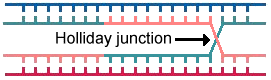
I. IntroductionHomologous recombination is essential for the generation of genetic diversity and for DNA repair in all organisms. A crucial step in recombination is the resolution of Holliday junctions, DNA intermediates which form as a result of strand exchange between two homologous DNA helices:
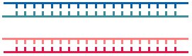
After the formation of Holliday intermediates, the length of heteroduplex DNA is extended through the process of branch migration. Heteroduplex double helical DNA contains two strands that were originally paired with other strands in the original double helices. Branch migration involves the sequential disruption of DNA base pairs in the two parental DNA helices, and the formation of new base pairs in the hybrid helices. Branch migration results in a region with two heteroduplexes and two original duplexes. To visualize this, it is useful to rotate the Holliday junction (the region near the arrow in the above figure) so that it has no crossing DNA strands and is in a square-planer configuration:
In Escherichia coli, branch migration and resolution of Holliday junctions to produce two, separate, intact DNA helices is accomplished by a suite of proteins: RuvA, RuvB, and RuvC. RuvA binds as a tetramer to the Holliday junction; this binding allows loading of RuvB, which acts as a molecular motor to drive branch migration. RuvC then cleaves the junction and resolves the two DNA molecules into mature recombination products. At left is shown an RuvA tetramer bound to DNA oligonucleotides representing a square-planer configuration of a Holliday junction. The regions of heteroduplex DNA (the top and bottom arms of the square planar DNA) correspond to the figures, above. II. RuvA StructureThe RuvA monomer has an "L" shape, composed of three domains: I, II, III. The loop connecting domain II and domain III is not shown. Domain I is a six-stranded, anti-parallel beta barrel, whereas both domains II and III consist entirely of alpha helices.
III. RuvA Tetramer-Holliday Junction DNAIn tetrameric RuvA, each monomer is a lobe of the symmetrical tetramer. The domain I beta barrels of each monomer are positioned centrally, and domains II and III are located peripherally. Examining the charge distribution of the surface of the RuvA tetramer, it is clear that the DNA binding surface is largely positively charged (basic), whereas the opposite surface is largely negatively charged (acidic). The basic nature of the "channels" that bind DNA attracts the negatively charged DNA backbone. An exception to the mostly basic surface of the DNA binding channels of the RuvA tetramer is an eight-residue acidic "central pin" containing glutamate55 and aspartate56 from each monomer. This negatively charged central pin may repel the negatively charged oxygens of the DNA backbone, driving the DNA away from the center of the Holliday junction.
IV. ReferencesAriyoshi, M., Nishino, T., Iwasaki, H., Shinagawa, H., Morikawa, K.: Crystal Structure of the Holliday Junction DNA in Complex with a Single Ruva Tetramer. Proc.Nat.Acad.Sci. 97: 8257-8262 (2000). Hargreaves, D., Rice, D. W., Sedelnikova, S. E., Artymiuk, P. J., Lloyd, R. G., Rafferty, J. B.: Crystal structure of E.coli RuvA with bound DNA Holliday junction at 6 A resolution. Nat. Struct. Biol. 5: 441-446 (1998). Roe, S. M., Barlow, T., Brown, T., Oram, M., Keeley, A., Tsaneva, I. R., Pearl, L. H.: Crystal structure of an octameric RuvA-Holliday junction complex. Mol. Cell 2: 361-372 (1998). |
|