THE BRAIN AND CRANIAL NERVES
Protected by Cranium
Surrounded by meninges and bathed in CSF.
Ventricles are CSF-filled chambers in the brain.
Lateral ventricle found within each cerebral hemisphere.
Ventricles
(Brazilian site: in English and Portuguese)
Ventricles:
QuickTime-VR clip, 3D
Ventricles:
3D cast, in situ
Ventricles:
3D cast, isolated
Images
from "David": Nice collection of images of meninges and ventricles
Brain
Ventricles & the Theory of the Mind: Historical Overview
Choroid plexus forms CSF --> CSF circulates thru ventricles and central canal of spinal cord. It reaches subarachnoid space via apertures and then flows thru the subarachnoid space of brain and spinal cord. Arachnoid villi absorb CSF and return it to the venous system.
Clinical Condition: Hydrocephalus ("water on the brain") if reabsorption of CSF is blocked in infancy. Expansion of ventricles causes thinning of cortex. Image.
Blood supply: Brain receives about the same amount blood at all times-- 20% of blood flow at rest; percentage falls during exercise but total flow increases so, little difference between rest and exercise in terms of flow.
High metabolism -- uses glucose for energy and doesn't store energy, so constant supply of glucose is necessary. High oxygen demand -- if interrupted, neuron death can occur rapidly.
Clinical Condition: Strokes (or cerebrovascular accident -- CVA)
Treatment: if stroke is due to an embolism, a "clotbuster" , e.g., TPA, may be used.
Aneurysm: thin wall of cerebral blood vessel expands and may rupture.
Two aspects of barrier function: 1: physical (capillary/astrocytes) 2:physiological (transport mechanisms)
Large molecules generally cannot pass from blood to the brain.
Capillaries are not very permeable. Lipid soluble materials get in easily (e.g., alcohol). Endothelial transport is very selective.
Regions of the Brain
Forebrain, Midbrain and Hindbrain -- found in early embryo.
These are further subdivided:
Forebrain:
Midbrain:
Hindbrain:
Cerebrum
Left and right hemispheres
separated by longitudinal fissure.
Superior
View of Brain
Fissures: deep gap between hemispheres and some lobes.
Corpus callosum -- tract that connects left and right sides.
"Split Brain" experiments of Sperry. Overview.
Segregation of some functions. Gender differences.
Gender
Differences and the Brain (see section on corpus callosum)
"Sex
and the Corpus Callosum" Summary
of lots of conflicting data on gender differences in the brain.
General Structure
Convolutions: consist of gyrus/gyri (fold/folds) and sulcus/sulci (groove/grooves). Function: increases surface area, complexity.
Example: Central Sulcus: Separates Frontal from Parietal Lobes. See pre- and post-central gyri below.
Lobes of Cerebrum:
Areas of Cortex (& relationship to Stroke)
"higher" thought, motor functions (voluntary).
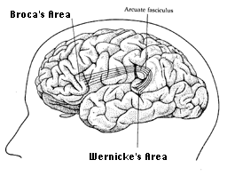
Broca's
area (speech-motor). Left side typically.
Broca's
area highlighted
Wernicke's area at junction
of occipital, temporal and parietal is involved in recognition of spoken and
written words.
Wernicke's
area - highlighted in light red
Connection between Wernike's
area and Broca's area:
Sensory (visual & auditory information related to language) is relayed to
the Wernicke's Area. If a word
is to be spoken, then the message will be sent to Broca's area.

Precentral gyrus -- motor representation of body.
Pre-motor Cortex: "fills in details" related to motor control. Sends input to primary motor cortex.
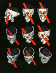
Phineus
Gage: Famous case history which demonstrated that part of our "social
brain" was localized to
the frontal lobe.
In mid 1800's there was a raging debate over whether specific functions resided in specific areas of the brain. Phrenology, a popular pseudoscience of that era centered around the belief that functions and personality traits could be discerned by examining bumps on the skull. Phrenologists had it partly right -- there is a localization of function to specific brain regions, but not skull regions!
Phineus
Gage Story in Discover Magazine
Phineus Gage Information Page Computer 3D reconstruction of Gage's injury (from Science Magazine)

Parietal Lobe: Sensory. Evaluation of general senses and taste.
Postcentral gyrus -- sensory representation of body.
Interpretative (Wernike's) area. Auditory cortex. Vestibular senses: motion, equilibrium & balance
Memory--short and long term memory integrated. Interface with limbic system.
Temporal lobe epilepsy can produce auditory hallucinations, feelings of deja vu, loss of sense of time.
Occipital
Lobe: Visual perception primarily. Each part of retina (visual space)
is represented by specific parts of primary visual cortex (area 17). Further
processing (interpreting images) is done in areas 18 and 19 of occipital lobe.
Receives input from retinas via the lateral geniculate nucleus, or LGN, of the
thalamus.
Simple Visual
Pathway.
Details
of Visual Pathway and Visual Fields
Limbic Lobe & limbic system:
Deep, medial portion of cerebral cortex (temporal lobe).
Insula: a "lobe" deep to temporal lobe. Part of olfactory cortex; connects with amygdala.Limbic system as a whole involves other areas of brain, particularly relating to memory and autonomic nervous system.
Regulation and display
of "emotions." A factor in autism?
Limbic
System and Autism
Area's of Brain Associated with "Emotion"
Hippocampus -- case history of H.M. (lesion resulted in "anterograde amnesia" -- he couldn't remember new things). Because of his case, researchers mistakenly believed that the hippocampus was the key place where conversion of short-term to long-term memory occured -- as always, it's more complicated than that!. The hippocampus and medial temporal lobe areas associated with converting short term into long term memory. Hippocampus does have a role in attention.
Amygdala: interfaces with sensory areas of other parts of brain; involved in memory. Recognition and display of fear.
Dr. Keele's "Amygdala Home Page"
Olfactory processing: Scientific American Article
Slide Presentation on Limbic System
White matter of cerebrum: large tracts. 3 classes
Diencephalon
Thalamus: primary relay center for sensory and motor pathways.
Thalamic Radiations: tracts going from thalamus to cortex
Dorsal View of Thalamus (purple); corpora quadrigemina below.
Sagittal View: thalamus, hypothalamus, pineal.
Thalamus (light blue); Hypothalamus (dark blue)
Hypothalamus:
Integrates autonomic nervous system -cardiovascular control, body temperature,
water/electrolyte regulation, hunger, sleep/wake cycles, sexual responses, control
of pituitary gland. Maintains homeostasis.
Remember the "4 F's": feeding, fighting, fleeing, fooling around
Area related to sexual
orientation? (Work
of Simon LeVay)
Homosexuality
and Biology (Atlantic Monthly article)
Epithalamus:
major structure is pineal gland (discussed in endocrine system). Produces melatonin
at night.
Salon
article on melatonin
Scientific
American article on melatonin
Mesencephalon (Midbrain):
Cerebral peduncles
"Corpora quadrigemina." 2 pair of colliculi ("little hills")
Superior colliculi: visual reflexes
Inferior colliculi: auditory reflexes
Sagittal View: colliculi, pineal, pons
Substantia nigra: motor activities. Affected in Parkinson's disease.
Metencephalon
Pons: relay station between medulla oblongata and midbrain. Origin of cranial nerves 5-8. Dorsal pons contains part of reticular formation (see below).
Cerebellum: lobed structure. Coordinate skeletal muscle contractions, muscle tone, balance, and timing. Integrates information from proprioceptors which monitor muscle and ligament tension, joint angles.
Cerebellum surface structure: note distinctive folia (rather than gyri) of cerebellum.
Anterior Lobe (Dorsal View, Cortex removed)
Vermis ("worm" - between left and right cerebellar hemispheres)
White mater: arbor vitae or "tree of life"
Sagittal View: cerebellum, pons, 4th ventricle
Anatomy and Physiology of Cerebellum: Site with very detailed Info.
Cerebellum overveiw Nice general site with links to other sites.
Myelencephalon
Medulla oblongata: at foramen magnum, becomes spinal cord. Similar to spinal cord except for "pyramids"--motor and sensory control tracts that run down spinal cord. Site of crossover (decussation) of left and right sides of body.
Medulla oblongata: sagittal view with pons and cerebellum
Medulla oblongata: anterior view showing pyramids
Origin of cranial nerves 8-12 (yes #8 is shared by Mesen- and Myelen-)
Control centers for respiration, heart beat and vasomotor control.
Brainstem = medulla, pons and midbrain.
Brainstem core contains reticular formation. Contains sensory (ascending) and motor (descending) pathways. Has centers for regulation of body functions. Lesion can result in coma.
Reticular activating system = the projections of the reticular formation. An "arousal" system. Sensory projections influence and regulate level of arousal and awareness -- sleep/wake cycles, motivation, levels of sensory perception, and emotions.
Human Brain: mid-sagittal section
Clinical Considerations:
Alzheimer's Disease: neurodegeneration with progressive accumulation of amyloid protein; "plaques" or "neurofibrillary tangles" form. Loss of memory. Genetic link.
Neurofibrillary
tangle: silver stain
Neurodegeneration
of cortex, Superior
& Lateral view
Loss
of cortex thickness results in expansion of ventricles
Epilepsy: Spontaneous
discharges (seizures) in different regions of brain.
Types
of Seizures: Petit mal, Grand mal and Psychomotor. Severity and form of
seizure dependent upon how much, and which part, of brain is affected.
Epilepsy
Foundation, Epilepsy
International
FDA
Report on Epilepsy
How
to Aid someone having a seizure
Cerebral Palsy: Quite variable since different regions of the brain can be affected. Motor disorder primarily, but mental retardation also occurs frequently. Can be bilateral or unilateral. Generally from prenatal or early postnatal cause -- trauma, low oxygen, strokes, meningitis. Rarely genetic.
Cranial Nerves: 12 pairs
1) Olfactory: Relays sensory
(olfactory) information from olfactory mucosa to olfactory bulb.
Olfactory
Nerve/Tract
2) Optic: Axons of cells from retina. Sensory (visual) information. Nerves cross at optic chiasma.
3) Oculomotor: Motor axons to most of the extrinsic muscles of eye and intrinsic muscles of iris--cause dilation of pupil. Some sensory info.
4) Trochlear: to superior oblique muscle. Trochlea means pulley or spool. Note sharp angle of superior oblique as it passes thru the trochlea of orbit.
5) Trigeminal: 3 branches--ophthalmic, maxillary and mandibular.
Tic douloureux: (Trigeminal Neuralgia) Disorder of maxillary and mandibular branches of Trigeminal. Very Painful. Can be treated with medication or surgery. Trigeminal Neuralgia Assn. , NINDS, newsarticle.
6) Abducens:
sensory and motor to lateral rectus.
(Neat
Graphic of extraocular muscles with abducens innervating the lateral rectus).
7) Facial: sensory and motor--facial expression and taste (front of tongue-sweet and salt), salivary glands of mouth.
Bell's
palsy: dysfunction of facial nerve. Inflammation or stroke results in
loss of function, for example facial
paralysis. Recent
studies have linked many cases to the "cold sore virus" a.k.a. herpes
simplex virus type 1 (HSV-1).
Other Bell's Palsy Sites:
NINDS,
MAYO, Bell's
Palsy Network
8) Vestibulocochlear (also called acuoutic or auditory nerve -- older terminology). Sensory primarily -- originates from semicircular canals (balance--vestibular sense) and from cochlea (hearing). Damage causes loss of hearing and/or problems with balance (vertigo).
9) Glossopharyngeal: motor to muscles for swallowing. Sensory from taste buds at back of tongue (bitter and sour).
10) Vagus: motor and sensory to viscera--heart, lungs, intestine etc. Carries some taste info.
Major nerve of parasympathetic nervous system.
11) Accessory: sensory and motor to trapezius and sternocleidomastoid.
12) Hypoglossal: sensory and motor to muscles of tongue (intrinsic. & extrinsic)
Ventral Veiw of Brain with labeled cranial nerves.
Inferior View of Brain: note cranial nerves
Cranial Nerves from "Neuroscience for Kids": Good, basic information!
LUMEN's Cranial Nerve Tutorial
Cranial Nerves - Yale School of Medicine (not complete yet, but some great info on many of the nerves)
Brain Comparisons
Comparative
Mammalian Brain Collection Homepage
List
of Specimens
Why are dolphins so darn smart?
Compare the brains of the flying fox bat (whose diet is primarily fruit) and that of the lesser horseshoe bat (whose diet is primarily beetles and moths). What structure related to their lifestyle is significantly different?
Human Brain Development
Miscellaneous Links
General Overview: Brain regions and functions
Gross Structure of Cerebral Cortex
Virtual Hospital's Human Brain Images
Harvard's "Whole Brain Atlas" MRI's of normal and diseased/damaged brains.
Anatomy/Phys Lecture Notes on CNS
U. Penn's Vet School Neuroanatomy (dog and cat brains)
Neurobiology at UC Davis Med School
UCLA's Laboratory of Neuroimaging Site has neat animations!
U. Utah WebPath's CNS Pathology Site
Neuroanatomy Lecture (Neuropsych course): good overview of cortical regions and functions.
Functional Anatomy of the Brainstem
Just for Fun