THE SENSES
Receptors are "transducers." They convert different energy forms to neural impulses.
Stimulus--> Receptor-->Impulse Conduction-->Interpretation
Sensation Vs. Perception
Classification of Senses
General and Special; Somatic and Visceral
Receptor Types
Exteroceptors:
Visceroceptors
Proprioceptors: e.g., muscle spindle, Golgi tendon organ.
Classification based on
Response and Adaptation
Phasic and Tonic Receptors
General Senses: Cutaneous & general sensations
Light touch - tactile corpuscles of Merkel and Meissner
Touch-Pressure - Pacinian corpuscle (lamellated)
Heat/Cold - free nerve endings
Pain - specialized free nerve endings. Respond to chemicals released by damaged tissue and to chemicals such as capsaicin which is found in chile peppers.
Capsaicin receptor: found on both pain and hot receptors.
Capsaicin Makes it Hot and Spicy: overview paper
Endorphins and enkephalins inhibit pain in brain.
Referred pain and Phantom limb
Proprioception - joint position, muscle tension
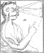
"5 Senses and Brain" image
Special Senses: Taste, Smell, Hearing, Equilibrium, & Vision
Smell - Olfaction
Based on Chemoreceptors.
Receptor cells are neurons. Rare example of neurons which undergo constant turnover in adult vertebrate nervous system.
Other Unique features of Olfactory Neurons and Olfactory Pathway
Embedded in nasal mucosa (olfactory epithelium). Fibers pass through cribiform plate of ethmoid bone and synapse in olfactory bulb. From there signals are sent to olfactory cortex of temporal lobe via olfactory tract. There are also connections to hypothalamus and limbic system (effects on emotions, memory, and reproductive cycles).
Transductive
Mechanism
Little was known of
olfactory transduction mechanism until recently. Involves molecules (odorants)
interacting with membrane receptors. Membrane receptors activate adenylate cyclase
--> cyclic AMP produced, which opens membrane sodium channel.
Overview site on Taste and Olfaction
"Pheromones," or air-borne hormones are detected by the vomeronasal organ (in nasal cavity but distinct from olfactory neurons). Pheromones are involved in mating, kin identification and bonding in many animals.
Olfaction and Human Kin Recognition
Vomeronasal Organ: Houses the receptors for pheromones
Taste (Gustation)
Chemoreception
Interacts with sense of smell.
Taste (gustatory) cells are housed in "taste buds" that are found along papillae of tongue. More taste buds
Taste bud anatomy - Histology 1, Histology2
4 types of papillae: fungiform, foliate, filiform (non-gustatory, mechanoreceptive), and circumvallate.
Histology: Circumvallate Papilla
Taste buds are embedded in epithelium of tongue. Communicate with outside via taste pores.
Cells are replaced on a daily basis. Turnover slows down with age.
5 Taste sensations and their localizations on human tongue:
However, the classic taste map of tongue is probably wrong!!!!!
Glutamate acts on other receptors which are found on other taste receptor cells; probably amplifies signals from all taste receptor cells. Monosodium glutamate acts as a "flavor enhancer" in this way.
Transductive mechanisms : The taste sensations are mediated by different transductive mechanisms.Taste is mediated by facia nervel (front of tongue) and glossopharyngeal nerve (back of tongue); vagus (palate/back of throadt) has a small role in taste.
Gustatory pathway -- nerves synapse in thalamus, relay to gustatory cortex of parietal lobe (tongue and mouth association areas of post-central gyrus).
There are differences, some which are genetic, in how people perceive tastes. For different tastes, one can be classified as a "non-taster," a "taster" or a "supertaster."
Overview of Taste: nice site.
Scientific American Article on Taste
Clinical Conditions
"Geographic Tongue" -- patchy loss of skin of tongue. Papillae lost.
Vision

Photoreceptors: Specialized hair cells which are adapted for capturing photons and generate an electrical response.
Structure of the Eyeball
Conjunctiva-- lines inside of eyelids and sclera that is exposed to outside. Secretes mucus. "Tears" are lubrication (from lacrimal glands). Tears also have anti-bacterial action. Histology
Chambers of Eyeball--Separated by lens and iris
Vitreous and Aqueous Chambers (with humors)
Vitreous humor is thick, gel-like consistency. Gives internal support.
Aqueous humor is more fluid and is found in 2 chambers: anterior & posterior.
3 Layers or Tunics of the Eye:
1) Fibrous tunic = sclera + cornea
Sclera = white of eyes. Cornea = clear region. Refracts light. Avascular -- oxygen must be taken in from air via moist surface.
2) Vascular Tunic = choroid, ciliary body--continuous with iris.
Iris-pigmented (color of eyes). Pupil regulates amount of light entering eye.
Lens--suspended to ciliary
body via suspensory ligaments.
Histology
Accomodation -- ability
to change from distance to near vision.
Contraction of ciliary muscle relaxes ligaments--> lens becomes more spherical--> near vision.
Relaxation of ciliary muscle contracts ligaments--> lens flattens--> distance vision.
Another cute demo of accommodation
Pupillary Reflex: pupils
dilate or constrict based on light intensity. Controlled by Autonomic Nervous
System.
Sympathetic: causes dilation of pupils. Parasympathetic: causes constriction
of pupil (via oculomotor nerve).
3) Retinal Layer
Simple
Overview
Labeled
Diagram, Basic
Cell Types
Retina: receptors and neural elements. An extension of the brain.
Pigment Layer (Retinal Pigment Epithelium). Pigment absorbs stray light and epithelial cells provide nourishment for photoreceptors (vitamin A transport) and takes away waste.
Retinal detachment -- Retina detached from pigment layer -- loss of nourishment leads to retinal damage and blindness.
Rods: 1 type-- for night or dim-light (black, white, gray) vision
Cones:
3 general types (red, green and blue)-- for day/color vision
Spectal
Sensitivities of Cones; Spectral
Sensitivities: WebVision
Distribution of rods and cones
Fovea centralis -- all
cones, all other neural elements moved away and no blood vessels overlying fovea
allows for less interference for incoming light. Histology
Monkey Fovea (labeled figure) Note lack of inner retinal cells in the fovea.
Macula lutea: circular, cone-rich area surrounding the fovea. "Lutea" refers to the yellow pigmentation which is present in this area.
Visual Transduction: Absorption of photons by photopigment (rhodopsin in rods) leads to hyperpolarization of photoreceptor -- photoreceptors are "turned off" by light.
Rhodopsin = opsin (protein) + retinal (derived from vitamin A, retinol). (Molecular structure of Rhodopsin)
Photon absorption by 11-cis retinal causes isomerization to all trans retinal --> this activates the opsin which, in turn, activates transducin which activates phosphodiesterase (PDE). PDE breaks down cyclic GMP to produce GMP.
cyclic GMP is what kept sodium/calcium channels open in the dark, so with a drop in cGMP concentration, channels close and photoreceptor hyperpolarizes, leading to a decreased release of neurotransmitters.
So, vertebrate photoreceptors are sometimes called "dark receptors" since they are active in the dark and turned off by light!
Another Visual Transduction Diagram
Photoreceptors synapse with interneurons (bipolar cells) which synapse with ganglion cells. Horizontal and amacrine cells are for lateral interactions.
Ganglion cell neurons--> axons become the optic nerve.
Optic disc--point where
ganglion neurons leave the eye to become the optic nerve--no photoreceptors
here -- basis of blind spot.
Blind
Spot Test
Histology
of Optic Disc
First Steps in Human Vision: General Overview on Access Excellence Web Site
Central Artery and Vein enter and leave via optic disc.

Optic
Pathway: optic nerves cross at optic chiasm (separation of left and right
visual fields),
optic tracts project
to lateral geniculate nuclei of thalamus and then to visual cortices of occipital
lobes.
Color Vision: Detailed info from "WebVision" site.
Opponent Colors & Color Afterimages
Optics:
Cornea and lens focus light on retina--image is inverted and shifted left to right. Shape of eyeball, cornea and lens dictate where light is focused.
Abnormalities:
Myopia: nearsightedness. Eyeball is too long--focus is in front of retina. Concave lens (or "minus") corrects.
Hyperopia: farsightedness. Eyeball is too short--focus is behind eye. Convex lens (or "plus") corrects.
Astigmatism: Corneal surface is irregular. Uneven lens required. Example Pattern.
Presbyopia: "elder eyes" -- loss of elasticity of lens and changes in suspensory ligaments occur with age, inability to close-focus--reading glasses (convex and magnifying) needed.
Refraction & the Eye Animations Site with downloadable animations covering refraction and eye disorders.
New Treatments: LASIK
Other Eye Diseases:
Color Blindness: hereditary, generally X-linked, red and green color vision deficits (males affected more than females). Blue-cone defects are more rare -- they effect males and females equally.
Color Blindness and Color BlindnessTests
Color Blindness: Sample Ishihara Plates
Just for Fun Colour Blindness Test
Night Blindness: Defective rod/peripheral vision. Can be from lack of vitamin A or beta-carotene --> insufficient production of retinal for rhodopsin. Gene causing one form of congenital stationary night blindness found recently. Review of CSNB.
Cataracts--cloudy lens due to direct or indirect damage. Lens normally yellows with age and cataracts are partly age related. There are some hereditary forms of cataracts.
Conjunctivitis: inflammation of the conjunctiva. From irritants or bacteria. Pinkeye is the bacterial form common in children.
Glaucoma:
increase pressure in eyeball due to poor drainage of aqueous humor -- damages
retina, esp optic nerve, if not treated.
Two major types - Open
angle and closed angle.
Retinal
Detachment:
retina separates from retinal pigment epithelium. Lack of nutrients results
in death of photoreceptors.
Excellent
site on Causes and Treatments.
Retinitis
Pigmentosa:
actually many different diseases. Inherited retinal disease (dominant, recessive,
and sex-linked forms). Rods affected first and fovea (cones) affected late in
disease. Tunnel vision. Mutations in rhodopsin or other rod-specific proteins
can lead to RP.
RP Q & A
Macular degeneration: affects macula (fovea and surrounding area which contains mostly cones). Age-related in part and influenced by pigment (more common in whites).
Diabetic Retinopathy: Diabetes results in capillary damage and edema. A leading cause of blindness in middle age.
Hearing and Equilibrium
Hearing: Based on Mechanoreceptors ("Hair Cells).
Structure of the Ear
Video Otoscopy Site: Lots of great images.
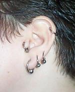
Tragus: A band and an region of the external ear!
External Ear
Auricle (aka pinna or helix) & External Auditory Meatus: Direct sound waves to Tympanic Membrane
Identify the pierced areas of the auricle!
 From: http://tattoo.about.com/library/weekly/aa041501b.htm
Meatus is lined with ceruminous
glands which secrete cerumen
(ear
wax). Functions: Waterproofing, inhibiting growth of micro-organims.
Accumulation/impaction of cerumen can be a problem
Meatus traverses part of temporal bone.
Middle Ear
Tympanic cavity within temporal bone. Separated from inner ear by oval and round windows. Connected to nasopharynx via Eustachian (auditory) tube. Functions to equalize pressure on both sides of tympanic membrane.
Contains Ossicles (little bones). Middle ear & Eardrum graphics.
Malleus (hammer), Incus (anvil) & Stapes (stirrup)
2 Primary Functions: Tranmission and Amplification of vibrations.
Two muscles dampen vibrations -- tensor tympani (malleus) and stapedius (stapes).
Inner Ear or "labyrinth"
Bony labyrinth outside, membranous labyrinth inside .
Perilymph separates bony and membranous labyrinth.
Endolymph within membranous labyrinth, surrounds sensory cells and structures.
Vestibule = utricle + saccule--gravity sensors, linear acceleration and deceleration.
Semicircular canals (3): angular acceleration and deceleration.
Cochlea: coiled, snail-like tube containing three chambers--upper and lower chambers (scala vestibuli and tympani), and a middle chamber (cochlea duct).
Sound = frequency (pitch -- cycles of compression per second) and intensity (loudness -- measured in decibels)
Mechanics of Sound Transmission
Organ of Corti is the sensory apparatus for hearing. Graphics & Histology
Range of human hearing:
20 - 20,000 cps or Hertz (Hz).
Higher frequencies
lost as we age
Hearing Range of Other
animals:
  Frequency Range of Hearing for Humans and Selected Animals

  animal                           frequency (hertz)
                                    low       high

  Humans                             20      20,000
  Cats                              100      32,000
  Dogs                               40      46,000
  Horses                             31      40,000
  Elephants                          16      12,000
  Cattle                             16      40,000
  Bats                            1,000     150,000
  Grasshoppers and locusts          100      50,000
  Rodents                         1,000     100,000
  Whales and dolphins                70     150,000
  Seals and sea lions               200      55,000
|
From: http://www.central-jersey-sas.org/projects/bat_detector/
Mechanism
Vibrations are transmitted to oval window. ---> Vibrations of oval window displace perilymph in s. vestibuli. ----> Endolymph is displaced by vestibular membrane ---> Force of perilymph transmitted to tectorial membrane of Organ of Corti. ---> Hair cells stimulated by tectorial membrane movement. ---> Sensory hair cells have output to auditory portion of vestibulocochlear nerve. Perception in temporal lobe.
Awesome Animations!!!! (Go to "Function")
Movement toward kinocilium depolarizes the sensory cell; movement away from kinocilium hyperpolarizes the sensory cell.
Tectorial Membrane helps to amplify movement of sensory hairs. (Cochlear Amplifier Animations)
High pitch is detected near base of cochlea, low pitch is detected further along the cochlea. Diagram and Animations
Length of sensory hairs of cochlear hair cells differs along the organ of corti -- short hair: high frequency; long hairs: low frequency.
SEM's of basal and apical hair cells: nice demonstration of hair length differences.
Cell length also differs and relates to frequency sensitivity.
Neurotransmitters of Organ of Corti
Diseases:
Conduction and Sensory Deafness: Numerous Types
Otitis
Media (common ear infection of children)
Meniere's Disease
(Washington Univ.)
Hearing Aids Before Electricity
Tinnitis: "Ringing in the Ears"
Ototoxicity: many chemicals are toxic to the inner ear.
Equilibrium
Receptor hair cells have kinocilia and stereocilia which are displaced by specialized structures.
Saccule and Utricle: Sensors of static equilibrium, gravity. They respond to linear motion of body.
Clusters of receptor cells (maculae) are embedded in jelly-like otolithic ("ear stones") membrane. Otoliths (also called statoliths or otoconia) are calcium carbonate crystals that are in otolithic membrane -- they provide inertia or drag. When head tilts, otoliths follow gravity. The gelatinous otolithic membrane moves and displaces receptor hair cells. Low Power Image
Semi-circular canals. They respond to changes in angular movement (rotation), called "dynamic equilibrium." 3 canals - one for each plane of the body. Each canal has an ampulla which contains a crista ampullaris (the structure which contains sensory cells). Sensory hairs (cilia) project into gelatinous cap-like cupula. Fluid movement in canal and ampulla displaces cupula and therefore the cilia are also displaced.
Crista ampullaris Histology, Histology 2, Histology 3. Histology 4 Hi Mag Labeled image
Neural pathway: Vestibular portion of vestibulocochlear nerve becomes vestibular tract which then sends information to brainstem, spinal cord, cerebellum, and cerebral cortex.
Nice Graphic of nerve & its relationship with other structures
Vestibulo-Ocular
Reflex
Animation
of Oculocephalic Reflex. (same as "vestibulo-ocular reflex above).
"Anatomy
of Petrous Bone" Nice CT / MRI views and reconstructions of inner ear.
Vestibular
Mechanisms Animations
Site with downloadable animations covering stereocilia movements.
Clinical Considerations
Inner Ear Infections can affect balance.
Motion sickness.
Miscellaneous Links
Sensory System
HHMI
"Seeing, Hearing and Smelling the World" (Howard Hughes Medical
Institute)
General Senses
Chronic
Pain Site
Taste & Olfaction
The
ChemoReception Web
Chemoreceptors
asscociated with taste and smell
Vision
WebVision:
Awesome site dedicated to the structure and function of the retina. (Note: this
site has been unaccessable recently)
Genetic
causes of retinal diseases
How
we see: Access Excellence
History
of Visual Science
Simple
Eye Test
Gross
and Microscopic Anatomy of the Eye
Visual
Illusions
Biology
of the Eye & Visual System: 14 powerpoint lecture slides
Color
Blindness & Web Design Site: examples of how dichromats would view color
photos.
Anatomy,
Physiology and Pathology of the Human Eye
Hearing/Balance
Virtual
Tour of the Ear
Vestibular
System Overview - nice graphics!
Auditory
and Vestibular Pathways
Lecture
Notes with mini-exam
The
Ear: Lecture w/diagrams