ARTICULATIONS
(JOINTS)
Chapter
10
An articulation or joint is any interface between two bones.
"Arthr-" is the common prefix or term relating to joints.
Kinesiology: the study of motion at joints and the interrelationships of bones, muscles and joints.
Historical Note: Hippocratic writings on joints
Classification of Joints
A) Structural Classes
B) Functional Classes (based on degree of movement)
Synarthroses--rigid and
immovable. Tight connection via fibrous tissue or cartilage.
3 subtypes.
Amphiarthroses--semi-movable.
2 subtypes.
Diarthroses -- freely movable
joints. Synovial joint enclosed within joint capsule.
Range of movement limited by bone, muscle and ligament attachments.
Structure: bones with articular
cartilages interface within joint capsule.
Synovial fluid between articulating surfaces. Articular cartilage is avascular.
Associated Structures of Synovial Joints:
Meniscus: cartilaginous
pads in joint capsule which serve as cushions and guides for articulations.
(See section on knee joint).
Bursa: sac filled with synovial fluid. Cushions muscles. Bursitis is an inflammation of bursa.
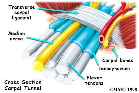
Tendon Sheath: modified
bursa-- facilitates gliding of tendons.
Example: Tendon
sheaths of wrist (carpal tunnel)
Bursitis:
emergency medicine site
Bursitis
- advanced info
Carpal
tunnel
Diarthroses Subtypes:
Movements at Diarthroses:
Angular Subtypes:
Circular Subtypes:
1) Rotation: no lateral displacement. E.g., at pivot & ball and socket joints.
At elbow: pronation &
supination.
At hip
and shoulder: medial & lateral rotation.
2) Circumduction: appendage
moved in cone-forming arc.
Distal end moved, proximal end is pivot. Shoulder, arm, trunk, hip and ankle.
Special Movements:
Overview of movements: Check out "Hypermuscle"
Jaw (Temporomandibular, TMJ)
Shoulder (Glenohumeral; Humeroscapular)
Elbow
Hip (Coxal)
Knee (Tibiofemoral)
Ankle (Talocrural)
Intervertebral
Southern California Orthopedic Institute: Excellent site covering major joints
Virtual Hospital’s site covering the major joints
Temporomandibular Joint or Jaw
Articulation between mandibular fossa of temporal bone and the condyle of the mandible.
1) Only movable joint in the skull
2) Combination hinge and gliding joint
Made possible by the articular
disk. Allows condyle of mandible to slide or glide forward and backward
(protraction & retraction) and laterally against the articular tubercle
of the temporal bone.
Articular disc separates joint into two regions.
Malalignment of TMJ results
in TMJ syndrome.
Condition is associated with pain in jaw, neck and shoulders along with ringing
in ear (tinnitis).
Jaw can easily be dislocated
-- mandibular condyle slides to far laterally or forward,
past normal location on articular tubercle.
Humeroscapular or Shoulder Joint
Multiaxial, Ball-and-Socket Joint. Most mobile joint of body.
Head of humerus articulates
in shallow glenoid fossa of scapula (although depth is increased by glenoid
labrum,
a fibrocartilagenous band around the glenoid fossa).
Allows flexion, extension, (rotation), adduction, abduction and circumduction.
High mobility due to:
Stability due to:
Baseball pitchers often have "rotator cuff injuries"-- this is when SITS muscles and/or their tendons are damaged.
The
American Shoulder and Elbow Surgeons Society
Shoulder
radiographs
Sports
Medicine and the Shoulder
Elbow Joint
2 uniaxial joints:
One joint capsule houses
both joints.
Medial
and lateral collateral ligaments reinforce joint.
lateral epicondylitis = "tennis elbow"
medial epicondylitis = "golfer’s elbow"
Elbow
anatomy - basic
Elbow
radiograph
Elbow
- regional
Posterior
Elbow Dislocation
Coxal or Hip Joint
Multiaxial, ball-and-socket joint.
Head of femur articulates in acetabulum formed by pelvic bones.
Compared with shoulder joint:
Greater strength and less mobility.
Like glenoid fossa, acetabulum is ringed by fibrocartilagenous lip or labrum.
Three major ligaments surround the outside of the joint and form the capsule. Ligaments arise from 3 bones of pelvis and attach to greater trochanter and neck of femur (thus, they are called iliofemoral, ischiofemoral and pubofemoral ligaments).
The teres ligament is present within the joint and attaches the head of femur to acetabulum.
Strength of ligaments of hip is so great that little muscular strength is needed to maintain standing position. But, walking with an upright posture means that hip joint must bear considerable loads during one's lifetime. Osteoarthritis of the hip is common.
Hip
Hip
joint - general
Hip
replacement site
Hippies
Hip
Hop
Tibiofemoral or Knee Joint
Largest and most complicated joint of the body. It is a modified hinge joint.
It is a composite joint
Functions of Patella: protection and leverage.
Basic motions are flexion and extension. Some rotation possible.
Main flexors are hamstring muscles, main extensor is quadriceps femoris.
Rotation is allowed by articulation
of round surface of femoral condyles with therelatively
flat surface
of tibial condyles. Medial articulation is wider than lateral articulation --
results in feeling that knee "locks into position."
Main ligaments:
The anterior cruciate becomes more taut with extension, prevents hyperextension and forward sliding.
The posterior cruciate becomes more taut with flexion to prevent backward dislocation and hyperflexion.
ACL
reconstruction
Arthroscopic
surgery to repair ACL
Meniscus (menisci, pl). Crescent shaped fibrocartilagenous plates within knee joint. Lateral and Medial
Functions:
Cushion between ends
of bones that meet in joint.
Fill space of non-matching
surfaces
Medial Meniscus is often
injured, esp. in football--it is attached to medial collateral ligament.
When knee is twisted, the ligament can tear the meniscus.
Torn
Meniscus
Arthroscopic
surgery to repair meniscus
"The 3 Cs of knee injuries"
Bursa in knee joints--around
patella.
"Water on the knee" is really frictional, or pre-patellar
bursitis
Talocrural or Ankle Joint
Two hinge joints involving three bones.
Distal End of tibia and medial malleolus articulate with talus.
Distal End of fibula and lateral malleolus articulate with talus.
Stability of joint is from malleoli and ligaments. (Deltoid ligament on tibial side; collateral ligament on fibular side).
Movements: dorsiflexion and plantar flexion.
Tendons (Achilles & extensor muscle) limit extent of movement.
(Inversion and Eversion entail ankle plus intertarsal joint movement)
Ankle
Rotating
foot and ankle
Fracture
of lateral malleolus xray
Intervertebral Articulations
The joints between vertebrae and intervertebral discs are considered cartilaginous, semi-moveable joints (amphiarthroses). There are synovial, gliding joints between the articular processes of adjacent vertebrae. Some movement is allowed between vertebrae and the vertebral column is stabilized by numerous ligaments and muscles.
As a whole, the spinal column can flex, extend and rotate.
Intervertebral discs: "hockey puck" shaped discs made up of a tough outer coat of fibrocartilage, the annulus fibrosus and a spongy internal nucleus pulposus. The nucleus pulposus is a remnant of the notochord, essentially the original "spine" of the embryo. The discs permit small movements and give resiliency to the vertebral column by acting as shock absorbers. A ruptured disc is when the nucleus pulposus breeches the annulus fibrosus.
Ruptured
disc
Ruptured
disc graphic
Ruptured
disc with laminotomy
Arthritis & Related Disorders
Rheumatism: any painful state of the supporting structures of the body.
Arthritis: a form of rheumatism in which is characterized by joint inflammation.
AMA’s Arthritis Site - good overview
HealthCentral Topic Center on Arthritis
Rheumatoid arthritis
Rheumatoid
Arthritis overview
Disfigurement
due to rheumatoid arthritis
X-ray
of hand with rheumatoid arthritis
Genetic
Differences in Immune System gene is related to severity of R.A.
Lyme disease
Bacteria triggers immune
reaction which is similar to rheumatoid arthritis. Carried by ticks (esp. deer
ticks).
Children with Lyme disease are often misdiagnosed as having juvenile rheumatoid
arthritis.
Lyme
disease description
CDC
page on Lyme Disease
Osteoarthritis
Osteoarthritis
overview
Osteoarthritis
of the spine - xray and gross specimen
Osteoarthritis
of the knee - xray
Osteoarthritis
of the hip
Osteoarthritis
of the fingers - xray
Gouty Arthritis
Gout
overview from New Zealand
Mayo
Clinic overview on Gout
Sodium
urate crystals from synovial fluid
X-ray
of foot with gout
Ankylosing Spondylitis
Ankylosing spondylitis resource page
Other Conditions Related to Joints
Dislocation: displacement of bone from joint; tearing of ligaments, tendons, and capsule.
Subluxation: partial dislocation.
Sprain: forcible wrenching/twisting of joint with partial rupture.
Strain: overstretching of muscle.
Kinesiology/Biomechanics Sites of Interest